

Dedicated solution for the analyses of cardiovascular MRI
Evaluating 100+ physiological parameters
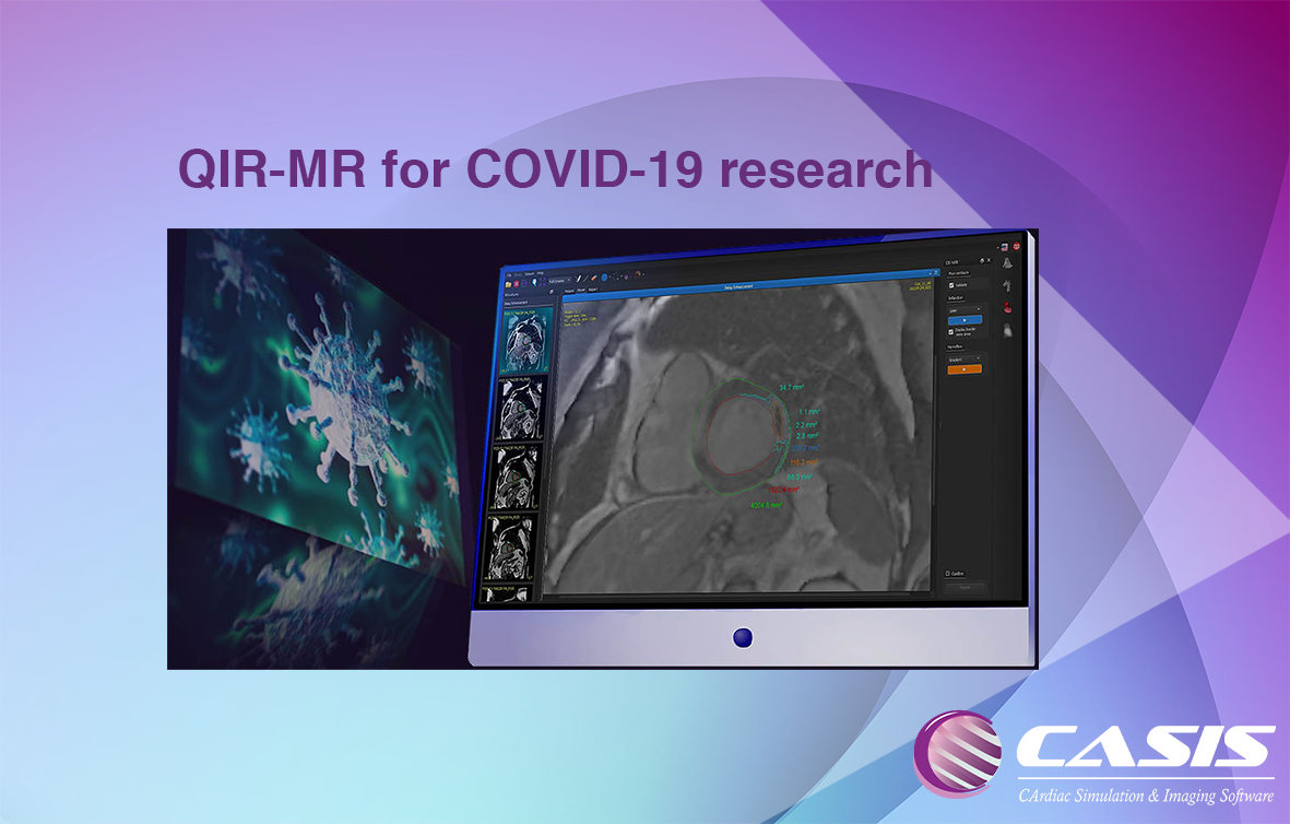
The virus COVID-19 can cause in some patients lesions of the myocardium visible in late enhancement MRI, a technique used to observe pathological areas of the heart
CASIS is offering free access to QIR-MR to all institutions studying COVID-19 and its effects on the heart
Our CMR software offers a set of modules, including Cine Cardiac, DE MRI, T1 and T2, to study heart conditions such as myocarditis and other myocardial injuries
QIR-MR is an improved software for MRI cardiac analysis. Functional analysis based on Deep Learning algorithm increases accuracy and speed of results. First Pass perfusion module assesses quantification of lack of gadolinium enhancement. It allows a better accuracy of area quantification between healthy areas and pathologic areas. Furthermore, clinical objectivity is improved. Quantification of Late Gadolinium Enhancement for each segment improves viability analysis and comparison between cardiac MRI exams. Original tools as aortic compliance and estimation of extracellular volume give advanced results for clinical practice and research issues. For clinical practice, the software ergonomics have been optimized with clinician’s feedback. Saving time is a priority for better work.
Benefits
WORKLOAD MANAGEMENT – Fully integrated solution in the workflow of the physician. Highly automated, with integration with PACS
PEACE OF MIND – QIR-MR contains the most clinically advanced algorithms providing anatomical guarantee
TIME SAVING – QIR-MR offers fast contour detection, resulting in less correction time. Therefore the physician can focus on the patient
Intuitive • Visual • Flexible
VIEWER
● AI-based automatic detection
● Flexible multisequence views
● Multi-screen advanced tab system
Common to all modules, the viewer allows you to:
● Intuitively navigate the software, with a simple interface
● Have a clear representation of all patient’s data. Display all loaded images of the exam in different viewers for better readability
● Work on different sequences simultaneously, and confirm the diagnosis with a complete view

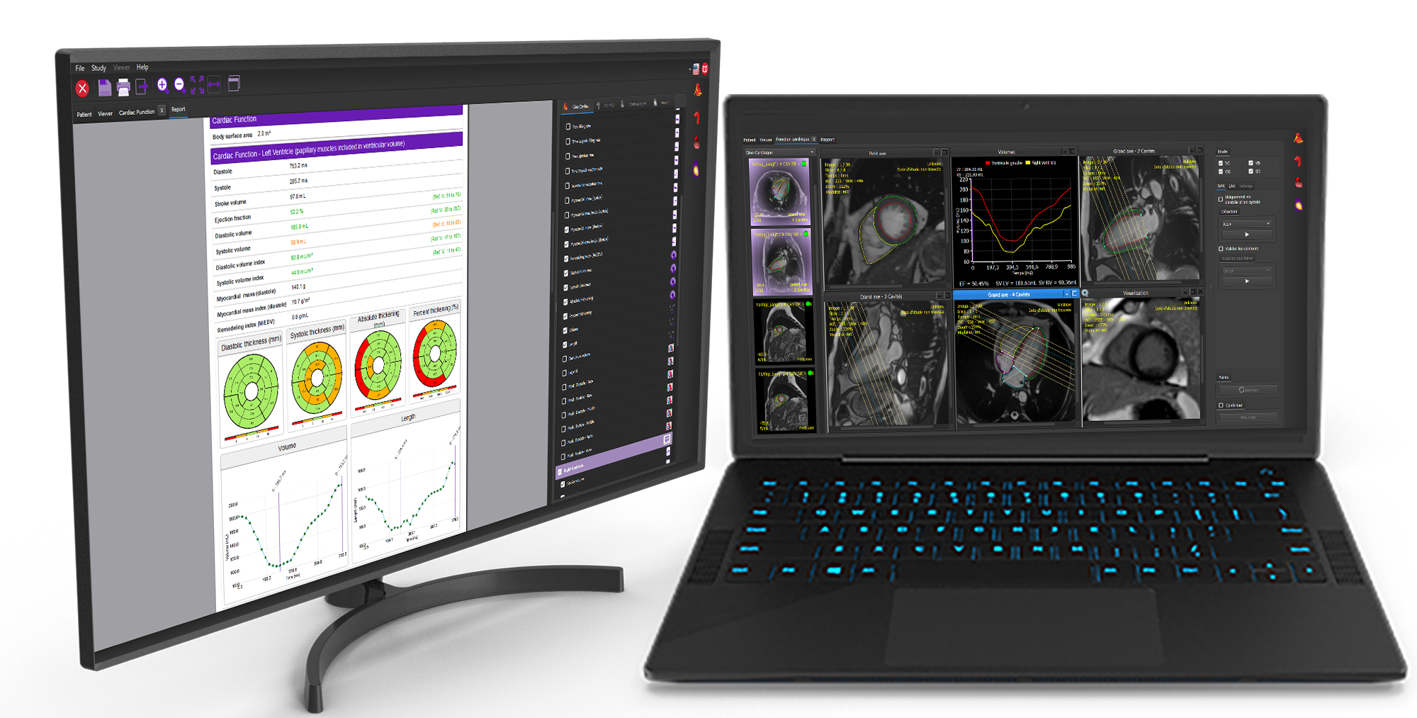
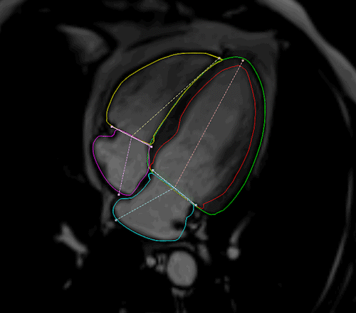
CINE MRI
Cardiac function:
- AI-based automatic detection
- Papillary muscle and trabeculation detection
- D/S SAX automatic detection option
- Comprehensive evaluation of ventricles and atria
- Contour detection of the 4 chambers based on AI
- User-friendly tools for intuitive contour adjustments
Strain:
- 4-chamber strain
- New methods: Fast and Simplified strain
- Radial, Circumferential, and Longitudinal strain
- Strain analysis using SSFP images
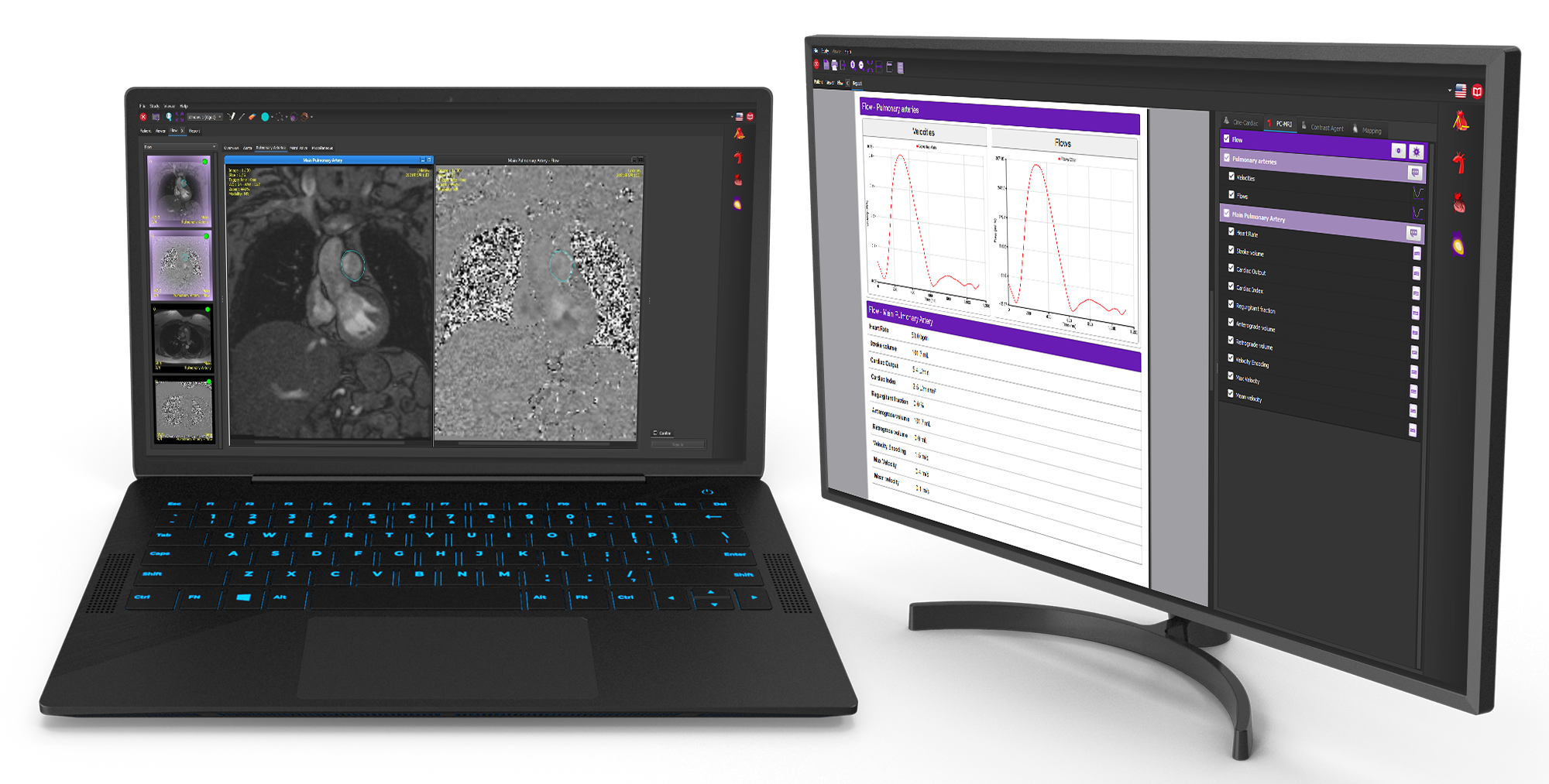
PC-MRI
Flow:
- 2D flow analysis
- Proximal vessels and mitral valve
- Pulse wave velocity and regurgitation fraction
-
Quantification (blood flow, regurgitation fraction, Qp/Qs)
Anatomical:
Assessment of:
- Aortic length
- Vessel’s compliance
- Aorta length and compliance assessment
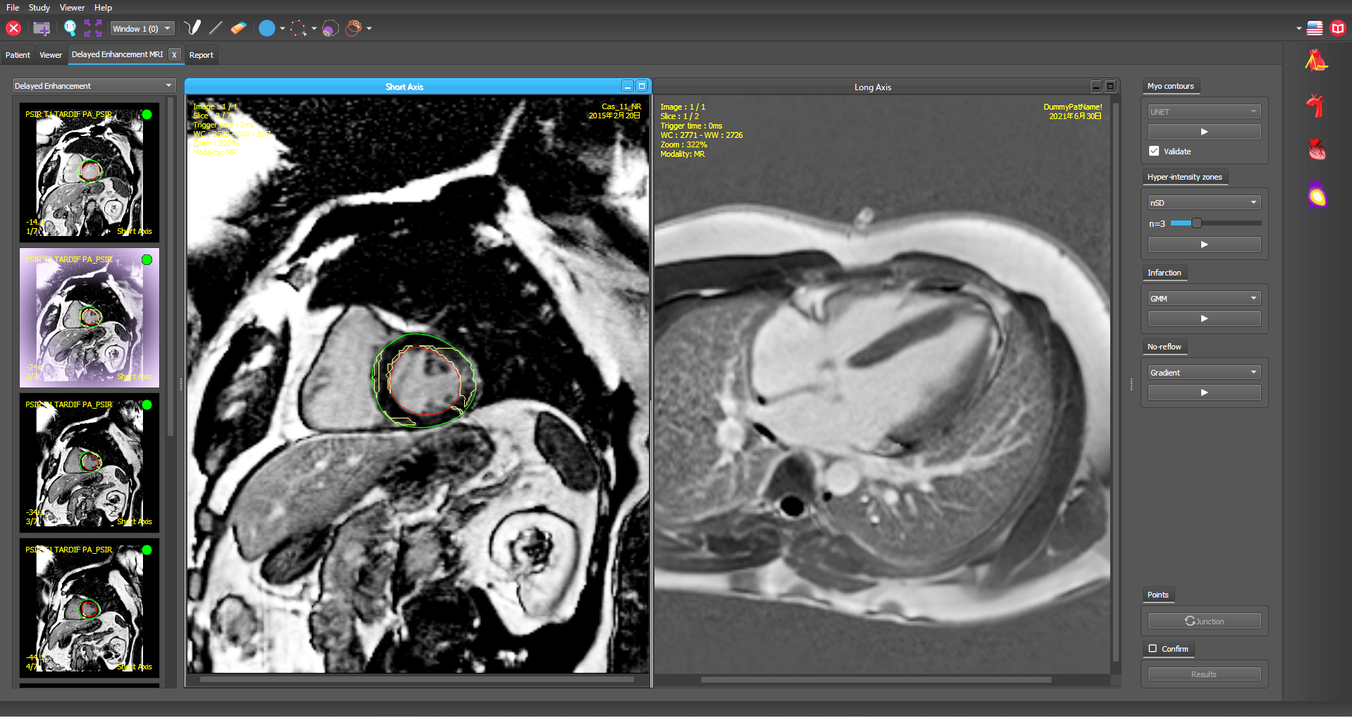
Contrast Agent
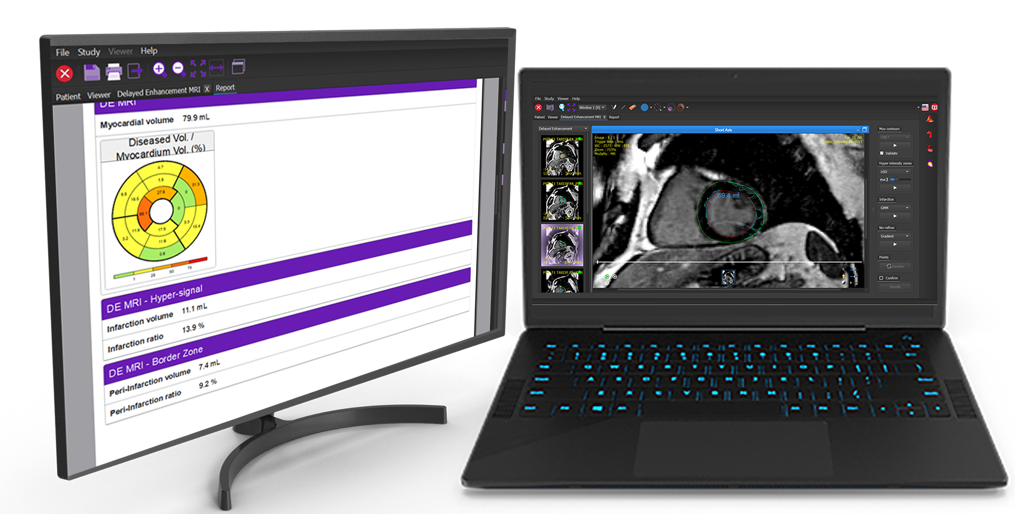
Delayed Enhancement:
- AI-based automatic contour and pathological area
- Characterization and quantification of pathological areas
- AI-driven LV contour detection
- Myocardial infarction : false-positive detection
First Pass:
- Stress vs. rest myocardial perfusion study
- Automatic contour propagation
- Motion correction
-
Perfusion signal intensity curve
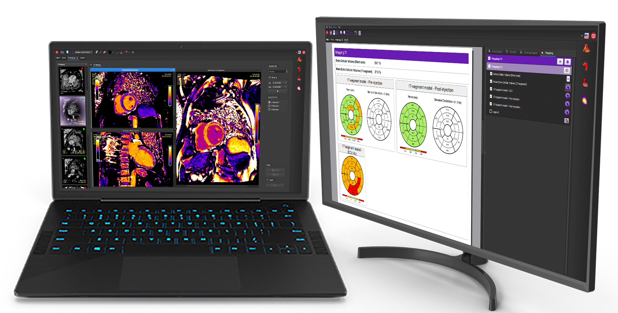
MAPPING
T1Mapping:
- Cartography reconstruction from native images
- Myocardium T1 and ECV assessment displayed for SAX and LAX
- Sorting of pre- and post- injection images
T2 Mapping:
- Myocardium T2 assessment
- Quantification displayed in 17/7 segments
T2* Mapping:
- Liver and myocardium T2* assessment
- Iron concentration evaluation
- Quantification displayed in 17/7 segments

Future
Is there something that you would like but haven’t seen?
Contact us and we’ll work with you to make it happen.